What does the oxide layer look like after a nickel-based superalloy has been oxidized at 1000°C for 100 hours? Under the Field Emission Scanning Electron Microscope (JSM 7800F), it appears as beautiful "peonies." Under the Nikon A1R confocal microscope, fluorescently labeled mesenchymal stem cells display their vitality like the splendor of a peacock's spread tail... These breathtaking microscopic photographs are works from a photography competition held at Tianjin University with the theme of Journey into the Microscopic World of Materials.
Since its launch in June 2024, the competition received 105 entries from 70 student groups across seven schools within the university. The competition enabled students to fully appreciate the unique charm of materials science.
Slected works
Theme: Peonies Sculpted by Time
Author: Cheng Xiaopeng from the School of Materials Science and Engineering
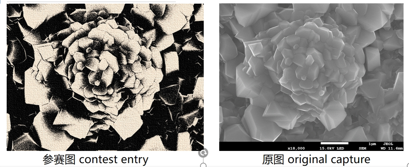
Description: The image, captured by a Field Emission Scanning Electron Microscope (JSM 7800F), reveals the surface oxide layer morphology after the GH3536 nickel-based superalloy was oxidized for 100 hours at 1000°C. During oxidation, spinel-structured oxide particles rapidly form on the sample surface. When colorized, these formations strikingly resemble a traditional Chinese ink painting of peonies. Under the influence of high temperature, these peony-like structures bloom across the sample surface, perfectly showcasing the traces sculpted by time.
Equipment Used: Field Emission Scanning Electron Microscope JSM 7800F
Theme: Flowering
Author: Cao Zhilong from the School of Materials Science and Engineering
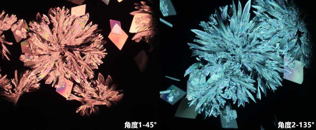
Description: The sample is a stimulus-responsive biomimetic liquid crystal elastomer material prepared through gradient cooling crystallization, which can be used to create soft robots for marine operations. The sample preparation process requires precise control of component ratios and reaction conditions. While the cooling rate must be very slow, the crystal growth occurs rapidly, making this visual effect fleeting and demanding exceptional timing to capture. The imaging requires precise adjustment of the polarizing microscope.
When rotating the specimen stage, the liquid crystals display different colors at various angles due to their birefringent properties. Here, red and blue colors were selected. The crystallization process of the liquid crystal resembles the blooming of flowers. In connection with its application in biomimetic robots, it's "as brilliant as summer flowers, a dazzling moment, like a fleeting flame across the horizon."
Equipment Used: Nikon Research-grade Polarizing Microscope Model: LV100NPOL
Theme: Colorful Lollipops
Author: Ye Jiamin from the Medical School
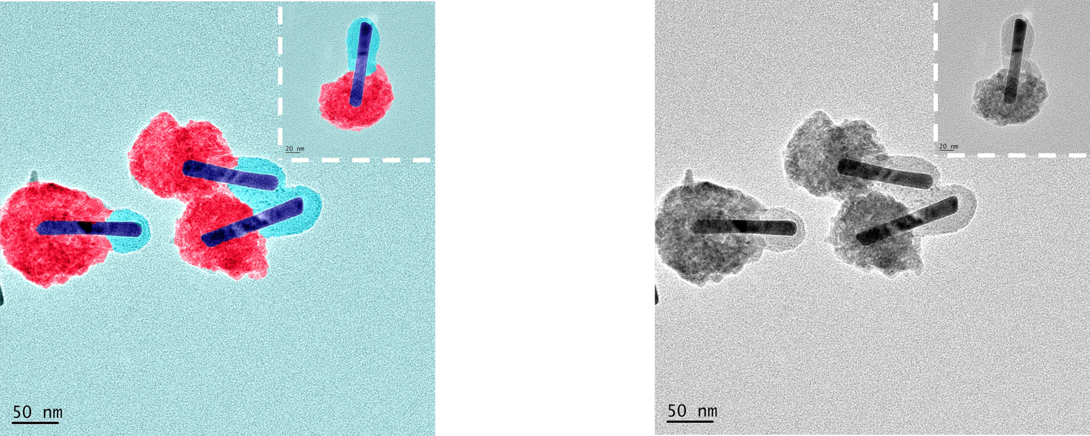
Description: This sample features a gold rod-silica-copper sulfide heterojunction material, synthesized in bulk through controlled wet chemical methods for potential applications in disease diagnosis and treatment. When colorized, the heterojunction material resembles colorful lollipops, characterized by one end being larger than the other. While research life can be challenging, the authors hope these "lollipop-like" materials bring a touch of "sweetness" to scientific work.
Equipment Used: Transmission Electron Microscope TecnaiG2F20
Theme: Peacock's dazzling tail
Author: Zhang Fanghua
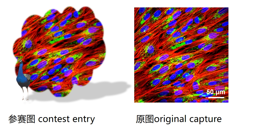
Description: This image, vibrant in color and rich in layers, resembles a peacock's spreading tail, symbolizing the splendor and vitality of life. The sample shows fluorescently labeled mesenchymal stem cells, used to study stem cell structure and function.
Equipment Used: Nikon A1R Confocal Microscope
Theme: Maple leaves
Author: Zhang Yang from the School of Materials Science and Engineering
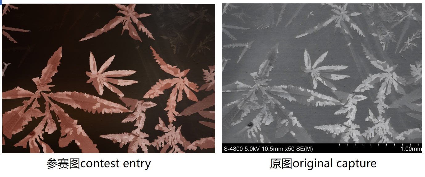
Description: The sample is a bismuth vanadate photoanode material loaded with flaky cobalt sulfate on its surface, used for photoelectrocatalytic water splitting. The preparation process requires precise control of the photo-assisted electrodeposition process.
After processing and colorization, the image evokes autumn's ripened maple leaves and fruits. The lighter surrounding areas resemble dappled light and shadows, while the deep background color suggests the vast, cloudless September sky.
Throught the works, the authors hope that students can possess the steady confidence of autumn days and the courage of maple leaves dancing in the wind, patiently awaiting the maturation of their research achievements - like maple leaves and fruits ripening - as they progress steadily toward abundant success.
Equipment Used: Scanning Electron Microscope S4800
By Eva Yin
